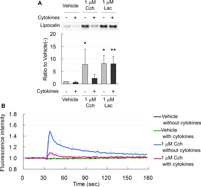
Figure 6.
(A) Representative immunoblot and band density measurements, showing that Lac and Cch equally induced lipocalin secretion from normal acinar cells not treated with cytokines (−). In acinar cells treated with 10 ng/mL each TNF-α and IFN-γ for 1 day (+), lacritin, but not Cch, induced secretion of lipocalin. Data are means ± SD (n = 6). *P < 0.05 relative to normal cells treated with vehicle and **P < 0.05 relative to cytokine-treated cells treated with vehicle. (B) Intracellular Ca2+ was increased by Cch in normal acinar cells (blue line) and was suppressed in cells treated for 1 day with 10 ng/mL each TNF-α and IFN-γ (red line).

