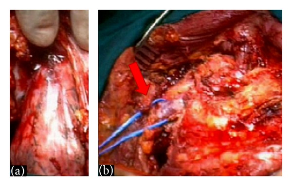
Figure 1.

Intraoperative view of a superior sulcus tumor: (a) The nervous and vascular structures lie above the tumor mass. With a correct surgical approach a posterior chest wall attached to round-shaped lesion was isolated from surrounding soft tissues. (b) The lesion was entirely removed without injury of the nervous and vascular structures. A full red arrow shows the integrity of the subclavian vein.
