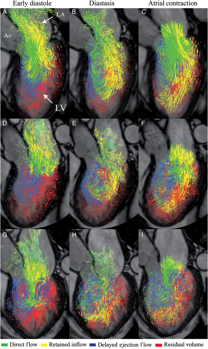
Figure 2.
Blood flow visualization: pathline visualization of the four flow components (direct flow, retained inflow, delayed ejection flow, and residual volume). (A–C) a healthy 50-year-old woman with normal LV diastolic function; (D–F) a 62-year-old male with DCM and LV relaxation abnormality; (G–I) a 61-year-old female with DCM and restrictive LV filling. Semi-transparent three-chamber images provide anatomical orientation. Ao, aorta; LA, left atrium; LV, left ventricle.

