Figure 3.

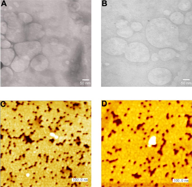
Morphology of the lipoplexes prepared at 10/1 (w/w) lipid-to-plasmid (p) DNA ratio. Transmission electron microscope images of a/pDNA (A) and b/pDNA (B); atomic force microscope images of a/pDNA (C) and b/pDNA (D).

Morphology of the lipoplexes prepared at 10/1 (w/w) lipid-to-plasmid (p) DNA ratio. Transmission electron microscope images of a/pDNA (A) and b/pDNA (B); atomic force microscope images of a/pDNA (C) and b/pDNA (D).