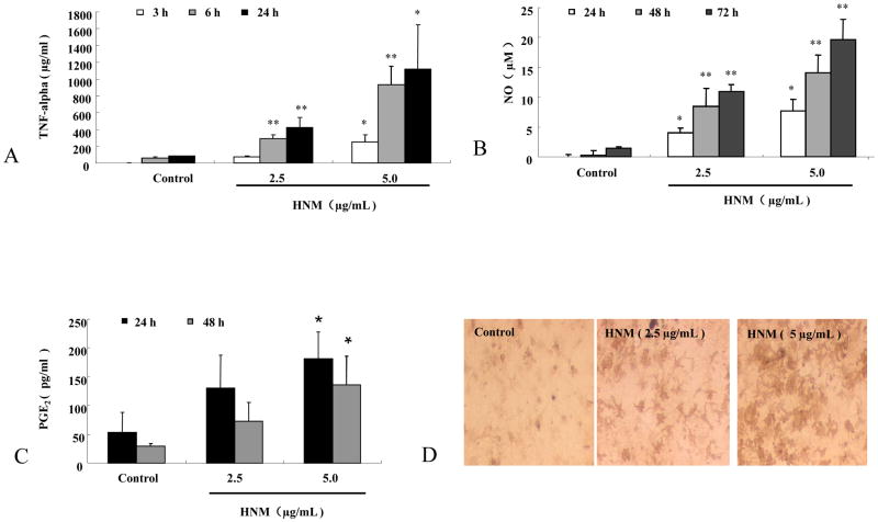
Figure 4. HNM increased the release of proinflammatory and neurotoxic factors in microglia.
Primary mesencephalic neuron-glia cultures were treated with 2.5 μg/mL and 5 μg/mL HNM, 50 μL supernatant was collected for the assays of TNF-alpha, NO and PGE2. TNF-alpha was measured 3 h, 6 h and 24 h after HNM treatment. *P<0.05, **P<0.01: HNM-treated group compared with vehicle-treated control group (A). NO was determined by measuring the accumulated level of nitrite (an indicator of NO) 24 h, 48 h and 72 h after the treatment. *P<0.05, **P<0.01: HNM-treated group compared with vehicle-treated control group (B). PGE2 was measured 24 h and 48 h after HNM treatment. *P<0.05: HNM-treated group compared with vehicle-treated control group (C). Representative microscopic images were shown for microglia treated with vehicle, 2.5 μg/mL and 5.0 μg/mL HNM, respectively, for 7 d (D).

