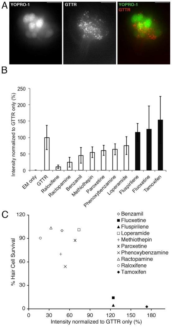
Figure 4.
Gentamicin-Texas Red (GTTR) entry into hair cells is altered by exposure to protective compounds. A) Images of a zebrafish neuromast pre-labeled with YOPRO-1 after a 3 min exposure to GTTR. YO-PRO1 labels hair cell nuclei (left, green channel). GTTR localized to puncta the apical region of hair cells, down and to the left in this image and a lower levels throughout the cytoplasm (middle, red channel). An image merging the red and green channels is shown to the right. The scale bar is 10 μM. B) Mean fluorescence intensity ± 1 s.d. of neuromasts exposed to protective compound and GTTR, embryo media only (EM only), or GTTR only (GTTR) for 3 min. Intensity is normalized and shown as a mean percentage of the GTTR only case. GTTR fluorescence intensity is significantly altered by addition of the protective compounds raloxifene (p<.001 ), ractopamine (p< .01), phenoxybenzamine (p<.05) and benzamil (p<.05). Shaded bars represent compounds that did not protect hair cells from gentamicin toxicity after 6 hr exposure. C) Plot of GTTR intensity following 3 min exposure to 50 μM GTTR (% intensity normalized to GTTR only) vs. hair cell survival following 6 hr exposure to 50 μM gentamicin (% hair cell staining) for each protective compound.

