
Figure 1.
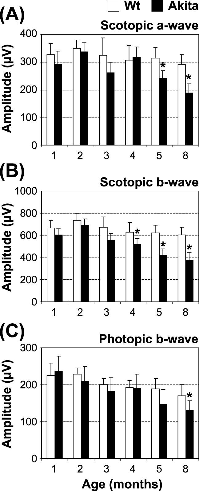
Functional analysis of the Akita mouse retina. Retinal function of Akita mice and age-matched wild-type controls was evaluated by ERG recording at the indicated age: scoptopic a-wave (A), scotopic b-wave (B), and photopic b-wave (C) are shown (mean ± SEM, n = 10, *P < 0.05).