
Figure 1.
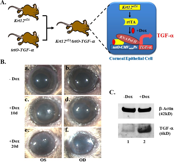
Overexpression of TGF-α in adult corneal epithelium caused corneal opacification and neovascularization. (A) Diagram shows the generation of bitransgenic mouse strain Krt12rtTA/tet-O-TGF-α. TGF-α overexpression in the corneal epithelial cells occurs upon Dox treatment. (B) Bitransgenic mouse eyes have clear cornea before Dox induction (−Dox, in [Ba, Bb]), but exhibited corneal opacity when treated with Dox for 10 days (+Dox 10d, in [Bc, Bd]). Noted blood vessels started growing toward the central cornea after 20 days of induction (+Dox 20d, in [Be, Bf]). (C) Western blotting revealed that the active form TGF-α (6 kDa) level increased dramatically after 10 days of Dox induction. β-Actin served as the loading control. OD, oculus dexter (right eye); OS, oculus sinister (left eye).