
Figure 3.
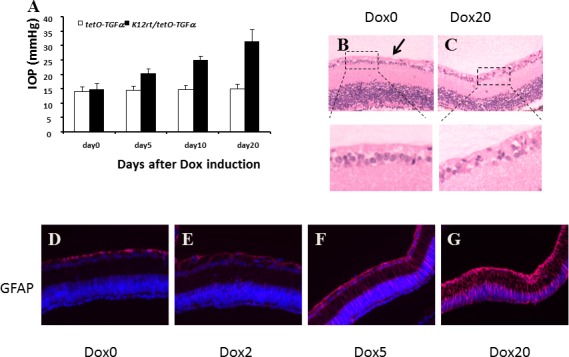
TGF-α expression led to elevated IOP, Müller glial cell activation, and retinal ganglion cell loss (A), IOP was measured in single- and double-transgenic mice before Dox induction and recorded as day 0. After 5, 10, and 20 days of Dox induction, IOP values of single-transgenic mice were steady during the course of induction, whereas IOP values of double-transgenic mice increased steadily and significantly. The data were collected from four mice of each group. TGF-α expression led to retinal ganglion cell loss (B, C). In control mice (B), the retinal ganglion cell layer consisted of one to two layers of well-organized cells (arrow). After 20 days of TGF-α expression, a single layer of sparse ganglion cells was evident (C). Immunoreactivity for GFAP in the retina revealed progressive activation of Müller glial cells by TGF-α expression (D–G). Before Dox induction (D), GFAP was localized in the nerve fibers and ganglion cell layer. After 2 days of induction (E), GFAP-positive signals were visible in all layers of the retina. After 5 days of induction (F), strong immunoreactivity was found in nerve fiber, ganglion cell layers, and in elongated cells extending across the whole section of the retina. The signal intensified after 20 days of induction (G).