
Figure 4.
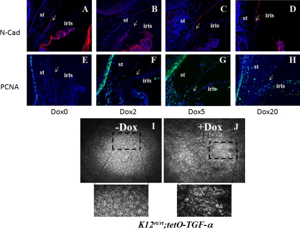
TGF-α expression led to corneal endothelial abnormality. N-Cadherin is the marker to track corneal endothelium. After 2 days of induction, corneal endothelium and trabecular meshwork were positive (A, B). After 5 days of induction, only the endothelium was positive (C). After 20 days of induction, N-cadherin–positive cells were also visible on the anterior iris, forming a continuous cell layer blocking the iridocorneal angle (arrow in [D]). Cell proliferation marker PCNA was positive in the corneal epithelium (E–H). Before induction, it was negative on corneal stroma and iris (E). Two to 5 days after induction, positive cells were also visible in the stroma (F, G). Twenty days after induction, both the corneal endothelium and peripheral iris were positive (H). Excess TGF-α changed the cell morphology and reduced cell density of the corneal endothelium. Live images of the corneal endothelium were taken by HRT II Rostock cornea module. Corneal endothelial cells mostly exhibited a hexagonal shape in untreated mice (I), but assumed diffuse abnormality with pleomorphism in size and shape in the treated mice (J). In addition, the endothelial cell density of Dox-treated mice reduced by 19.4% as compared with the nontreated mice (from 2736 ± 28 cells/mm2 to 2204 ± 43 cells/mm2).