
Figure 5.
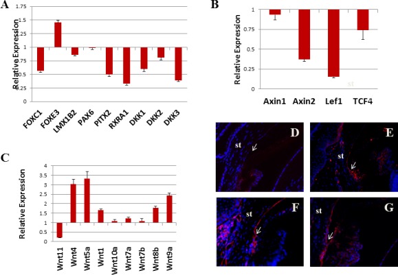
Noncanonical Wnt signal was activated in TGF-α–expressing cornea. Real-time qRT-PCR revealed that Pitx2, RXRα, and DKK3 were downregulated more than 2-fold following 10 days of Dox induction compared with uninduced control (A). Canonical Wnt targets (Axin2 and Lef1) were also downregulated (B). Noncanonical Wnt ligands (Wnt4, Wnt5a, and Wnt9a) were upregulated (C). Noncanonical Wnt target, MLC-P, was weakly positive in uninduced corneal endothelium (D). Two days after induction, trabecular meshwork and nearby endothelial cells showed stronger signal (E). After 5 days of induction, positive cells can be seen in both the trabecular meshwork and corneal endothelium (F). After 20 days of induction, positive signal can be seen on both sides of the iridocorneal angle (G). st, stroma; arrows point to iridocorneal angle.