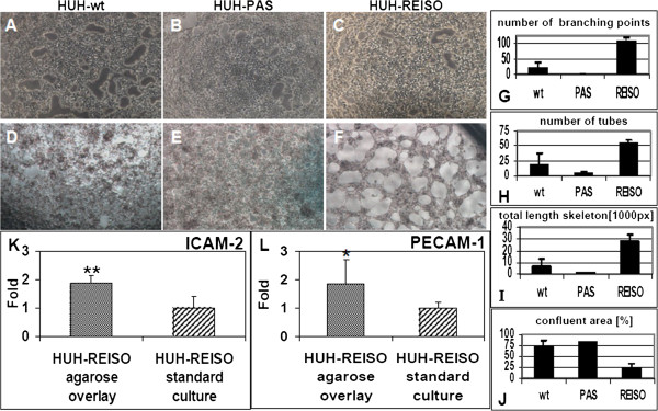
Figure 9.

Tube formation assay. Tumor cells were seeded on matrigel after conventional cell culture (21% Oxygen / 37°C) or after a 42 days culture in a diffusion limited environment with reduced oxygen and nutrient supply. Pictures were taken 24h after plating. (A) Parental HUH7 (HUH-wt), (B) in vivo passaged (HUH-PAS) and (C) chemoresistant (HUH-REISO) cells derived from conventional cell culture did not show tube formation, whereas in the case of cells, derived from the diffusion limited environment, the (F) chemoresistant tumor cells (HUH-REISO) showed significant tube formation potential in the matrigel assay, in comparison to (D) HUH-wt and (E) HUH-PAS (n=5). Software based analysis of pictures revealed a significantly increased (G) number of branching points, (H) overall number of tubes and (I) total length skeleton in chemoresistant cells (HUH-REISO) in comparison to both control cell lines (HUH-wt and HUH-PAS). Consequently, (J) the total confluent area was significantly decreased for HUH-REISO (n=3). A qRT-PCR analysis, performed after the tube formation assay on the endothelial markers (K) ICAM-2 and (L) PECAM-1/CD31, revealed significantly increased expression levels for cells derived from the diffusion limited environment culture compared to conventional cultured cells, which did not show tube forming potential (n=6).
