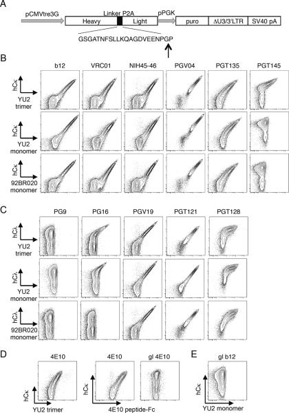
Figure 2.
Design and expression of bNAb antigen receptors by lentivirus transduction. (A) Schematic arrangement of vector showing gene elements encoding antibody genes and adjacent elements. The H-chain genes carry a leader exon followed by VDJ codons and constant region for membrane IgM. This is followed in frame by the picornavirus P2A peptide, shown below, and the light chain VJ and C codons. Also shown is the puromycin resistance gene (puro). The vertical arrow shows the P2A “cleavage” site, which lacks a peptide bond upon translation. (B–E) Expression and soluble Env binding by the indicated bNAb BCRs in transduced WEHI231 cells. Final concentration of antigens used in staining was 25 μg/ml, except for 98BR020 monomer, which was at 20 μg/ml.

