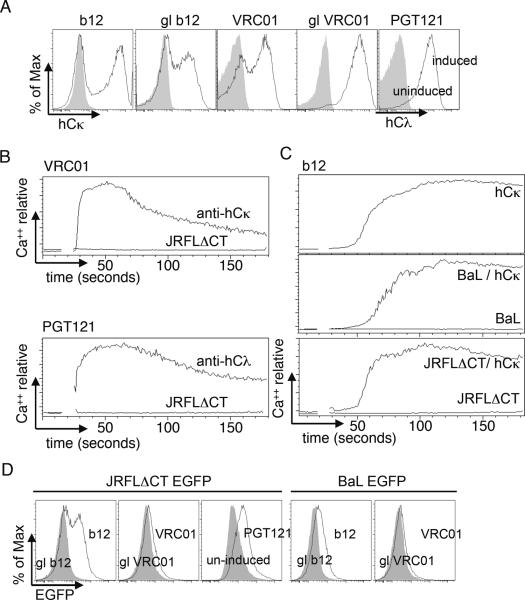
Figure 3.
Analysis of inducible expression and the ability of GFP-labeled pseudovirions to activate bNAb-expressing WEHI231 cells. (A) Comparison of human L-chain expression in induced (black line) versus non-induced (grey fill) cells carrying the indicated bNAb genes. (B,C) Ca++ mobilization analysis of the indicated bNAb-expressing cell lines upon stimulation with anti-hCκ (10 μg/ml) or the indicated pseudovirions (100× concentrated stock final concentration, approximately 3×10⁹/ml for JRFLΔCT and 2×10¹²/ml for BaL; the JRFLΔCT concentration used corresponded to 1 nM Env trimer equivalents as established by ELISA). (C) b12 cells were stimulated with anti-hCκ alone (top panel). In the lower two panels activation by the indicated pseudovirions (lower traces), was followed by challenge with anti-hCκ (upper traces). (D) Analysis of pseudovirion binding in the same experiment, using the same virus particles and concentrations as in B and C, but incubating on ice for 1 hour. Filled traces shown binding to uninduced cells or cells carrying germline reverted (gl) bNAbs.

