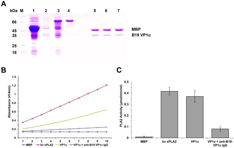
Figure 1. Purified VP1u proteins and PLA2 activity.
(A) SDS-PAGE analysis of expressed fusion protein and the purified protein VP1u or mutants cleaved by Factor Xa digestion. M: Protein marker; lane 1: Total protein of E. coli DH5α transformed with pMAL-c2x with IPTG induction; lanes 2 and 3: Total protein of E. coli DH5α transformed with pMAL-VP1u with or without IPTG induction, respectively; lane 4: Purified fusion protein MBP-VP1u; lanes 5, 6, and 7: Purified VP1u, VP1u-H153A, and VP1u-D195A digested by Factor Xa. (B–C) PLA2 activity on various substrates as indicated at the bottom. The PLA2 activity is shown as a value of the absorbance at 414 nm at various time points. Purified MBP, 5 µg; bee venom, 10 ng; purified VP1u (5 µg); VP1u (5 µg) with anti-B19 VP1u (5 µg). Data are representative of three independent experiments.

