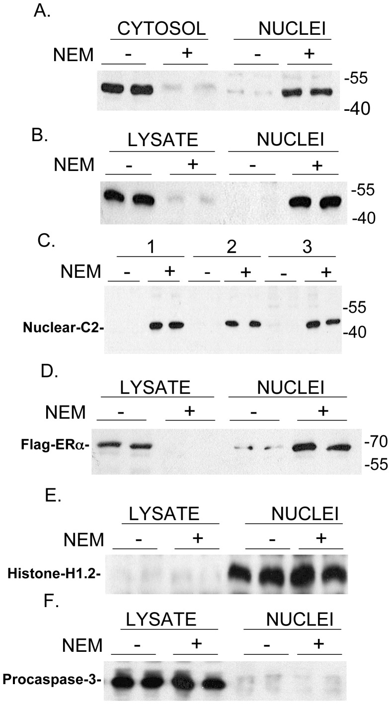
Figure 1. Effect of N-Ethylmaleimide (NEM) on the distribution of endogenous caspase-2 (C2) (Panels A–C) and FLAG-ERα (Panel D) between nuclear and extra-nuclear (cytosol or lysate) cell fractions.
(A) Caspase-2 in cytosol and nuclei of cells without (−) or with (+) NEM (20 mM) pre-treatment for 10 min prior to cell fractionation using hypotonic buffer without detergent. (B) Caspase-2 in the lysate and nuclei of cells without (−) or with (+) NEM (7.5 mM) pre-treatment for 10 min prior to cell lysis with buffer containing 0.5% Triton X-100. (C) Nuclear Caspase-2 in cells without (−) or with (+) NEM (7.5 mM) pre-treatment for 10 min prior to lysis using the following conditions: 1) after lysis in cell culture plates (lysis buffer added directly to the plate wells), 2) cells in suspension collected in an Eppendorf tube were instantly frozen in dry ice/ethanol, transferred to ice bath and ice-cold lysis buffer with Triton X-100 was immediately added to the frozen pellets, 3) cells in suspension collected in an Eppendorf tube at room temperature and room temperature lysis buffer with 0.5% Triton X-100 was added to the pellets. (D). FLAG-tagged ERα in the lysate and nuclei of cells without (−) and with (+) NEM (20 mM) pre-treatment for 10 min prior to cell lysis with buffer containing 0.5% Triton X-100. The numbers on the right of the panels reflect the gel migration of the 40 kDa, 55 kDa, and 70 kDa protein markers. To ensure that our cell lysis procedure actually reflects nuclear and extra-nuclear (LYSATE) fractions, Western blotting studies examined for Histone H1.2 as a nuclear marker (E) and Procaspase-3 as an extra-nuclear marker (F) [31]. Cell lysis was carried out using the conditions given in (B) above.

