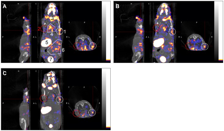
Figure 6. In vivo imaging of Bac-NIS-infected hUCB-MSCs transplantation with NanoSPECT/CT.
A: Overlapping SPECT and CT images at 30 min after administration of 300 µCi (11.1 MBq) Na125I. The right axilla (white circular area) was transplanted with Bac-NIS-infected hUCB-MSCs (MOI = 200, 1×10⁷ cells) and showed a high radioiodide uptake, while the left axilla (red circular area) was transplanted with mock-infected cells as a control and showed no obvious radioiodide uptake. From left to right side are, respectively, the coronal, sagittal and horizontal sections. All CT images are shown with a grey palette, and all SPECT images are shown with a warm palette. B and C: SPECT/CT images at 60 min and 120 min after radioiodide administration. Abbreviations: CT, computed tomography; SPECT, single photon emission computed tomography; 1, Bac-NIS-infected hUCB-MSCs; 2, mock-infected hUCB-MSCs; 3, thyroid; 4, heart; 5, stomach; 6, intestinal area; 7, bladder.

