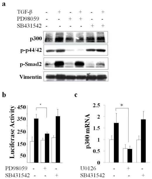
Figure 3. p300 stimulation by TGF-β is mediated via ERK1/2.
a. Confluent fibroblasts were preincubated with SB431542 (left panel) or PD98059 (right panel) followed by TGF-β2 (12.5 ng/ml) for 24 h. Whole cell lysates were examined by immunoblot analysis. b. NIH3T3 fibroblasts transfected with p300-luc were preincubated with SB431542 or PD98059, followed by TGF-β for 24 h. Whole cell lysates were assayed for their luciferase activities. Open bars, untreated fibroblasts; closed bars, TGF-β-treated fibroblasts. The results normalized with protein concentration represent the means ± S.D. of triplicate determinations. * p<0.05. c. Confluent normal adult dermal fibroblasts were preincubated with SB431542 or U0126 followed by TGF-β2 for 24 h. The real-time qPCR results, normalized with GAPDH, represent the means ± SD of triplicate determines from the representative experiment. *, p<0.05.

