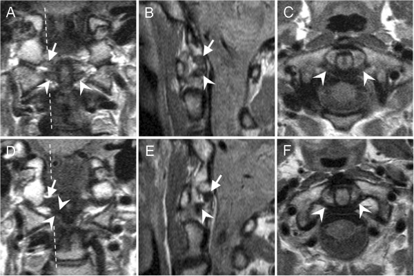
Figure 1.

Scoring high signal intensities of alar and transverse ligaments on upper neck MRIs. Proton-density-weighted, fast-spin echo, 1.5 Tesla MRI sections were performed in (A, D) coronal, (B, E) sagittal, and (C, F) axial directions. MRIs were from two healthy women, aged (A-C) 44 years old, and (D-F) 60 years old. Broken lines mark the sagittal plane. (A-C) The transverse ligament is indicated with arrow heads. The high intensity signal was scored 2 by reader A, 1 by reader B, and 2 by consensus; in the second evaluation, the same signal was scored 2 by both readers independently. The alar ligament is indicated with arrows. (A, B) The high intensity signal was graded 2 by both readers independently; in the second evaluation, the same signal was scored 2 by reader A, 3 by reader B, and 2 by consensus. (D-F) The transverse ligament (arrow heads) and alar ligament (arrows) were scored 0 by both readers independently in both evaluations.
