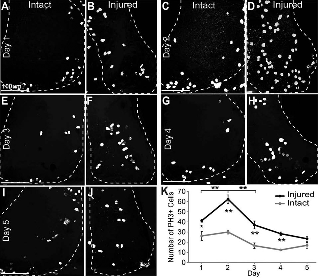
Figure 4. Phospho-Histone 3 immunoreactivity increases after injury.
The right tectal lobe of stage 47 tadpoles was injured and changes in cell division were assessed in the right and left tectal lobes by immunolabeling for phospho-histone H3 (PH3) to identify dividing cells in M phase of the cell cycle over the course of 5 days. Data were analyzed as individual optical sections, but are presented as confocal Z-projections of the tectal lobes, which are outlined. A–J. PH3 labeling of dividing cells in the tectal lobes 24hrs (A,B), 48 hrs (C,D), 3 days (E,F), 4 days (G,H), and 5 days (I,J) after injury in the intact left tectal lobe (A,C,E,G,I) and in the injured right tectal lobe (B,D,F,H,J) of the same animals. K. Total counts of PH3-labeled nuclei in the injured (black line) and intact (gray line) tecta over a 5-day period after injury. N=25 animals total (5 for each time point). *p<0.05, **p<0.01, n.s. = not significant. Scale bars, 100 µm. Data are average ± SEM.

