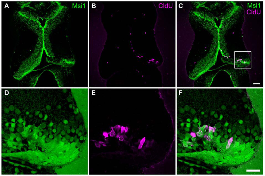
Figure 6. Proliferating cells are musashi1-expressing neural progenitor cells.
Stage 47 tadpoles were injured in the right tectum. Two days later animals were exposed to CldU for 2hrs and processed immediately for CldU immunolabeling. A–F. Representative single optical sections from confocal images of 30 µm sections through the optic tectum of tadpoles labeled with antibodies to musashi1 (Msi1, green) and CldU (red). D–F show enlargements of the boxed area marked in C. 93.3 ± 2.5% of CldU-positive cells in injured tectal lobes were double-labeled with musashi1 antibodies (n=7 animals). Note that the injury is restricted to the tectal cell body layer and the neuropil remains intact.

