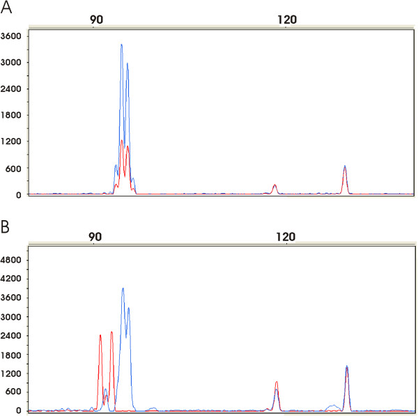
Figure 3.
Analysis of EGFR expression in non malignant and tumor colorectal tissues using fluorescent multiplex RT-QMPSF. After adjustment on peaks corresponding to control genes (PGK and SF3A, peaks on the right), amplicons from normal (in blue) and tumor (in red) tissues are superimposed. A: Expression profiles in a non mutated sample from a patient with A13/A14 genotype. B: Expression profiles in a mutated sample from a patient with A13/A14 genotype; notice in the tumor sample a shift of the peaks to the left corresponding to A9 and A11 repeats.

