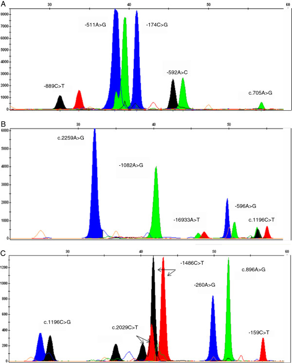
Figure 1.
Electropherograms obtained for the three multiplex primer extension panels. Capillary electrophoresis of SNaPshot products was performed with an ABI 3130 Genetic Analyzer. (A) Panel 1 SNaPshot results obtained for IL1-α: rs1800587 (−889C>T), IL1-β: rs16944 (-511A>G), IL6: rs1800795 (−174C>G), IL10: rs1800872 (−592A>C) and IL12: rs11575934 (c.705A>G). (B) Panel 2 SNaPshot results for TLR2: rs5743708 (c.2259A>G), IL10: rs1800896 (−1082A>G), TLR2: rs4696480 (−16933A>T), IL6: rs1800797 (−596A>G) and TLR4: rs4986791 (c.1196C>T). (C) Panel 3 SNaPshot results for IL12: rs401502 (c.1196C>G), TLR2: rs121917864 (c.2029C>T), TLR9: rs187084 (−1486C>T), CD14: rs2569190 (−260A>G), TLR4: rs4986790 (c.896A>G) and CD14: rs2569191 (−159C>T). Nucleotides are represented by the following colours: A = green; C = black; G = blue; T = red.

