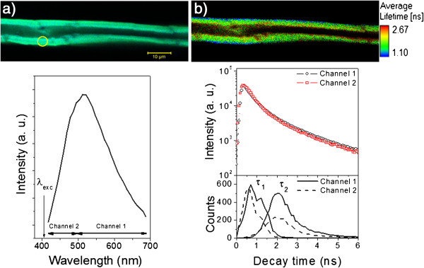
Figure 1.
Confocal and FLIM images for a single bagasse fiber: spectral and fluorescence decay features. a) Spectral confocal image of a single bagasse fiber treated with NaOH 0.5% excited at λexc = 405 nm (continuous wave). The spectrum below corresponds to the emission evaluated at the yellow spot of the cell wall. The spectral regions for channels 1 and 2 used in the decay time measurements are displayed in the figure. b) The corresponding FLIM image and the associated decay features detected from channels 1 and 2. The figure below shows the decay time distributions for τ1 and τ2 evaluated from the FLIM image for channel 1 (solid lines) and channel 2 (dashed lines).

