Fig. 4.
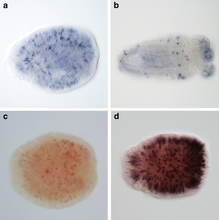
In situ hybridization (ISH) expression patterns of NEP-16 in Nematostella. a Three-day-old planula. b Seven-day-old primary polyps. Note the distinct long nematocyte-like ectodermal cells in the body wall and tentacle tips (dark blue staining). c Single NEP-16 expression stained with FastRed. d Double ISH of NEP-16 and the nematocyst marker NvNCol-3 (dark brown–purple) show perfect localization of NEP-16 to nematocysts. In all pictures, the oral end of the animals lies to the right

