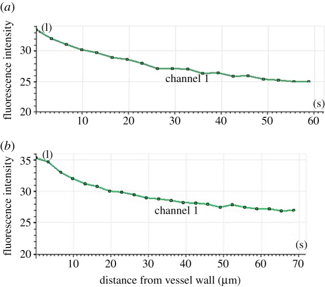
Figure 5.

LDL distribution on the y-axis of the rabbit carotid artery as determined by LSCM. The hyalinized rabbit carotid artery was perfused with medium containing DiI–LDL. The distribution of LDL concentration in the artery was measured along the z-axis using LSCM. (a) Half (cupular part) and (b) the other half (basal part) of the blood vessels in the analysis chart.
