
Figure 7.
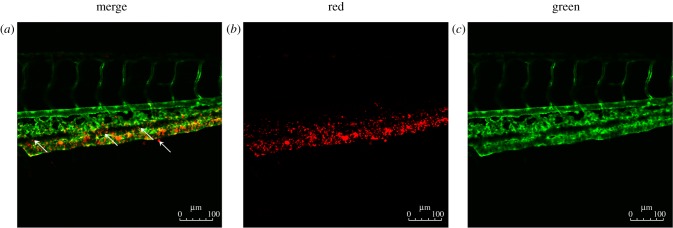
LDL accumulation and permeation in the vascular system. (a–c) Flk1: GFP embryos at 72 h.p.f.; embryos injected with Dil-LDL at 48 h.p.f. (n = 56/60); endothelial cells are green and Dil-LDL is red. Arrow denotes location of LDL permeation.
Official websites use .gov
A
.gov website belongs to an official
government organization in the United States.
Secure .gov websites use HTTPS
A lock (
) or https:// means you've safely
connected to the .gov website. Share sensitive
information only on official, secure websites.
LDL accumulation and permeation in the vascular system. (a–c) Flk1: GFP embryos at 72 h.p.f.; embryos injected with Dil-LDL at 48 h.p.f. (n = 56/60); endothelial cells are green and Dil-LDL is red. Arrow denotes location of LDL permeation.