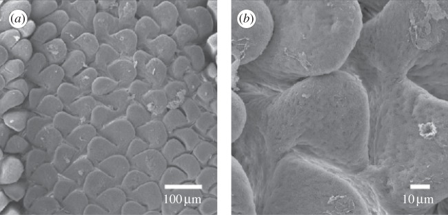
Figure 2.

Scanning electron micrographs of emerging villi in the jejunum of turkey embryos (reproduced with permission from [4]). The micrographs are taken at 21 days of incubations, and shown using scales of (a) 100 μm and (b) 10 μm for outlining of the morphology of the bi-dimensional undulated pattern at the free surface of the mucosa.
