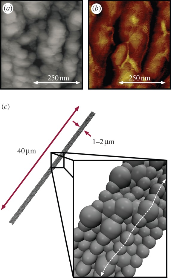
Figure 2.

Experimental and model silk fibre cross sections. (a,b) Experimental images of fibril structure in the core region of dragline silk fibres. (a) Topographic AFM image of spider dragline silk from Nephila pilipes, from Du et al. [7], used with permission, copyright 2006 Elsevier Inc. (b) AFM image from Nephila clavipes (golden orb weaver), from Brown et al. [41], used with permission, copyright 2012, American Chemical Society. Both images depict braided bundles of fibrils on the hundreds of nanometre scale constituting the fibre's core, with irregular ‘globular protrusions’ along the fibre axis. Moreover, small voids were observed in the images, indicating potential free volume for hydration. (c) Model silk fibre consisting of aligned and bundled fibrils with random globular protrusions to mimic the observed natural structure. Here, 20% of the particle sites are designated ‘globules’ (with a diameter of 300 nm) and randomly distributed along the fibril length, with an equilibrated diameter of approximately 1.2 μm. (Online version in colour.)
