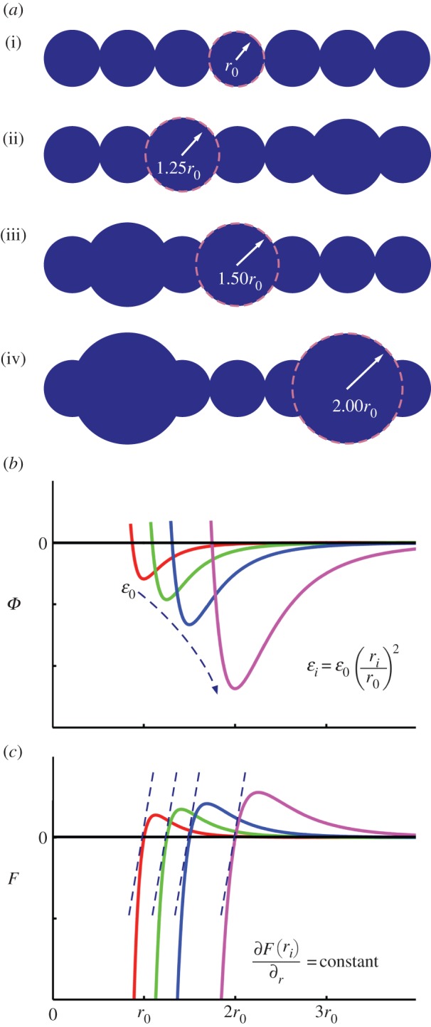
Figure 3.

Fibril computational model. (a) Depiction of relative particulate size along the modelled fibrils, including (i) homogeneous fibril (r0; 0%g), and three globule sizes (ii) rg = 1.25r0, (iii) rg = 1.50r0 and (iv) rg = 2.00r0. The globules are randomly distributed amongst all fibrils in the fibre model, with %g ranging from 5% to 50%. (b) Plot of energy versus separation for LJ 9 : 6 potential for fibril contact (ϕcontact) depicting scaling of parameters: as rg increases, the LJ parameter scales with ɛi ∝ (ri/r0)2 to maintain constant stiffness. (c) Plot of force versus separation for LJ 9 : 6 potential, depicting equivalent small deformation stiffness for r = r0, 1.25r0, 1.50r0 and 2.00r0. (Online version in colour.)
