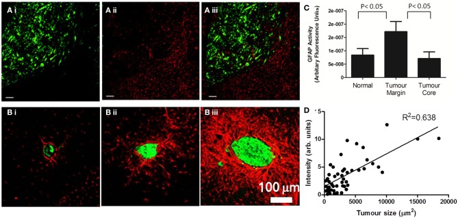
Figure 1.
Astrocyte activation, as determined by GFAP staining, is present in the peri-tumoral area of both primary (A) and secondary (B) tumors. (A) (i) In a mouse model of glioma, the growth of DBRTG glioma cells can be seen in green (GFP labeled), adjacent to a wall of astrocyte activation, seen in red (Alexa555 probe) (ii). The merged image (iii) indicates little infiltration of activated astrocytes into the tumor mass, as quantified by arbitrary fluorescence units of GFAP activity (C). Figure adapted from Lee et al. (2011). (B) In a mouse model of lung derived brain metastasis, in which HARA-B cells were inoculated intra-cardially, astrocyte activation (Cy3 probe, red) was observed surrounding metastatic growth (Alexa488 probegreen). As with the glioma model, astrocytes are present in the tumor periphery, rather than the core. The extent of astrocyte activation increased with tumor size (D). Figure adapted from Seike et al. (2011).

