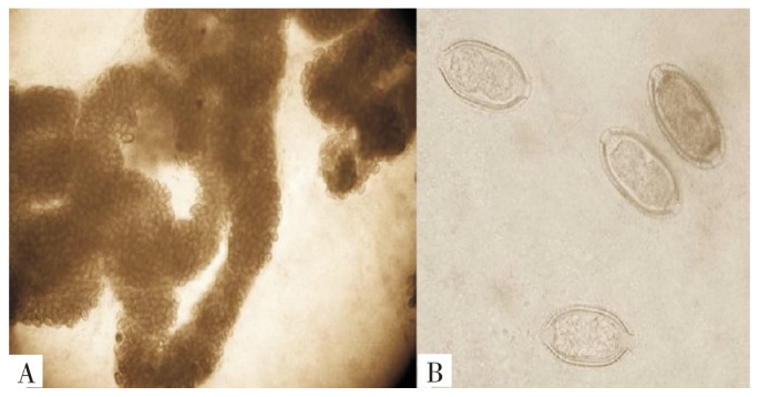
Figure 1. Morphology of C. hepatica eggs in the liver of the infected rats (unstained).

A, low-power view of the eggs in worms in pressed preparation liver tissues (100×). B, high-power view of the eggs. Note the characteristic barrel-shaped bioperculated eggs showing a thick shell with striation of the outer layer ( 400×).
