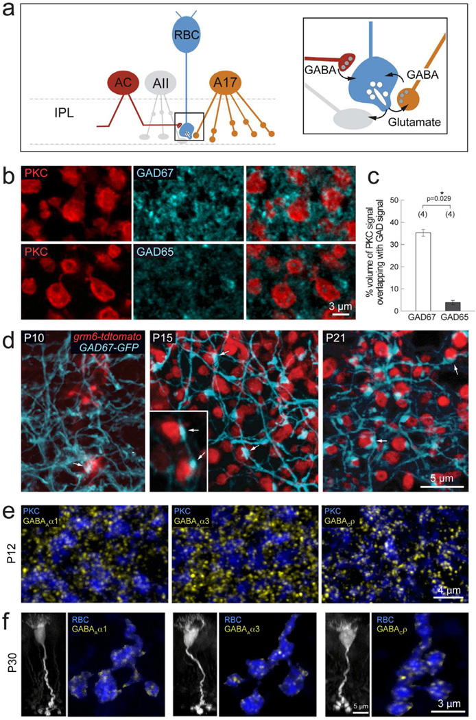
Figure 1. Development of GABAergic synapses onto RBC axon terminals.
(a) Schematic showing RBC connections with amacrine cells in retina IPL. RBCs (blue) provide excitatory glutamatergic drive (glu) onto glycinergic AII (grey) and GABAergic A17 (orange) amacrine cells. In turn, they receive feedback inhibition from A17 cells and additional GABAergic input from other as yet unidentified amacrine cells (AC, red). (b) PKC labeled RBC terminals (red) colabeled with GAD67 or GAD65 (cyan), reveal more abundant immunoreactivity of GAD67 as compared to GAD65 in the IPL lamina where RBC axons stratify. (c) Quantification of % volume of GAD signal overlapping with PKC immunoreactivity revealed a significantly higher % overlap with GAD67 positive processes as compared to GAD65. Asterisk marks significant difference. (d) Development of contacts (arrows) between RBCs (red) and GAD67-GFP positive amacrine processes (cyan) in the grm6-tdtomato×GAD67-GFP double transgenic line. By P15, large varicosities can be seen at sites of contact between RBC axon terminals and GAD67-GFP amacrine processes. (e) Axonal terminals of P12 PKC-labeled RBCs (blue) express GABAAα1, GABAAα3, and GABACρ receptor clusters (yellow), indicating the presence of these three GABAergic postsynapses on RBC terminals before eye-opening. (f) P30 RBCs in the grm6-tdtomato transgenic line (left, vertical view, grayscale). Axonal terminals of individual P30 RBCs (horizontal view, blue) express all three GABA receptor cluster types: GABAAα1, GABAAα3, and GABACρ (yellow),

