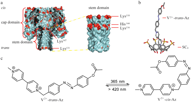
Figure 1. Representation of an α-HL (PDB ID: 7AHL) and the interaction mechanism between SC4 and V2+-trans-Az.
(a) An α-HL nanopore is embedded in a lipid bilayer at the Tris-EDTA (pH 8.0). The two compartments of the bilayer cell are termed cis and trans. The potential is applied through Ag/AgCl electrodes and the cis compartment is defined as virtual ground. Left: Cross section of α-HL which consists of a large cap domain at the exterior of the membrane and a transmembrane stem region exhibiting a diameter of 1.4 nm in its narrowest constriction. Right: the illustration of outer surface of stem domain. His, Arg and Lys as positive-charged residues are coded in red. Lys110, Lys116, Lys131, His144 and Lys147 are positioned at the stem of α-HL, respectively. (b) The representation of SC4:V2+-trans-Az complex. V2+-trans-Az is immersed into the cavity of SC4 in its axial orientation with the viologen group being included first. (c) The photoisomeric reaction of V2+-trans-Az to V2+-cis-Az upon irradiation with UV light at 365 nm.

