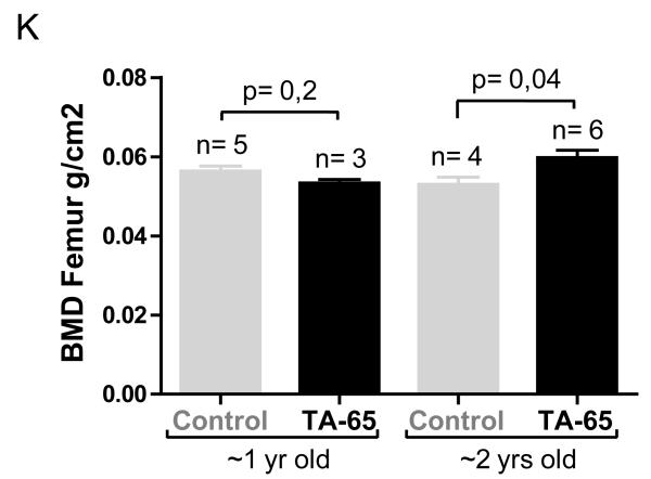
Figure 5. TA-65 treatment delays some age-associated pathologies.
A. Thickness of the subcutaneous fat layer at time of death in the different mice cohorts. B. Representative images of subcutaneous fat and epidermal layers in mice feed with vehicle or vehicle plus TA-65. C. Thickness of the skin epidermal layer, at time of death, in the different mice cohorts. D. Stimulation of wound closure in HEKn cells incubated in the presence of TA-65. Images were taken after incubating for 24, 48 and 72 hours. Wound closure was calculated and is presented in the right panel. E. Hair re-growth capacity was quantified in arbitrary units (a.u., see Materials and Methods) 14 d after plucking. Fisher’s Exact test was used for statistical analysis. Experiments were carried 12 months post the ending to the TA-65 supplementation period in the 1 yr old cohort of mice. Six independent mouse were used. F. Representative images of hair-regrowth. Images were acquired in anesthetized female mice of the 1 yr old group before and 14 days after hair plucking. G. Percentage of Ki67-positive cells in the epidermis (tail skin) from mice non-treated (control) or treated with TA-65. Student’s t-test was used for statistical assessments. At least 6 high-power fields (HPF, x100) were used per independent mouse (around 2000 skin epidermis cells scored per mouse). H. Representative Ki67 immunohistochemistry images of skin epidermis (tail) from the indicated mice cohorts. I. Percentage of TUNEL-positive (Apoptag detection kit) cells in the epidermis (tail skin) from mice non-treated (control) or treated with TA-65. Student’s t-test was used for statistical assessments. At least 6 HPF (x100) were used per independent mouse (around 2000 skin epidermis cells scored per mouse). J. Representative TUNEL stained images of skin from the indicated mice cohorts. K. Femur bone mineral density (BMD femur) measured at the time of death in the 2 yrs old cohorts.


