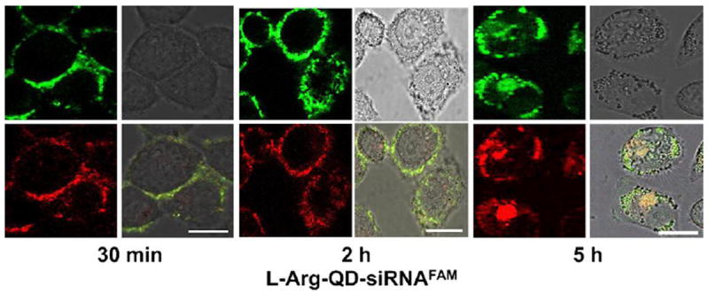
Figure 8.

Real-time confocal microscopy images of HeLa cells transfected with CdSe/ZnSe QD/siRNA complexes. Soon after transfection, the QD/siRNA was observed on the cell membrane. At 5 h, QD/siRNA had migrated into the cell cytoplasm. Green fluorescence represents siRNA and red fluorescence represents QDs.
Figure with permission from Li, J.-M., M.-X. Zhao, H. Su, Y.-Y. Wang, C.-P. Tan, L.-N. Ji, and Z.-W. Mao, Multifunctional quantum-dot-based siRNA delivery for hpv18 e6 gene silence and intracellular imaging. Biomaterials, 2011. 32(31): p. 7978-7987.
