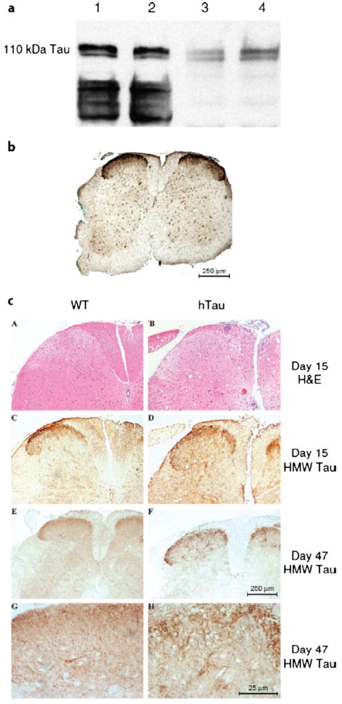
FIGURE 3.

The 110-kDa high-molecular-weight (HMW) tau is expressed in the spinal cord during experimental autoimmune encephalomyelitis (EAE). (a) Immunoblot analysis for HMW tau in mouse spinal cord. Twenty micrograms of protein homogenate from the lumbar spinal cord of wild-type (WT) mice at 2 months (lanes 1, 3) and 9 months (lanes 2, 4) of age was loaded per lane. Lanes 1 and 2 were incubated with mAb tau 46 (1:1000); lanes 3 and 4 were incubated with HMW tau polyclonal antibody (pAb) (1:20,000). (b) Spinal cord section from a mouse with high levels of human tau (hTau) stained with HMW tau pAb (1:10,000). (c) Paraffin-embedded sections of WT (A, C, E, G) and hTau (B, D, F, H) mice stained with hematoxylin and eosin (A, B) or immunostained with anti-HMW tau polyclonal antibody (C–H). Scale bars = (b, c [A–F]) 250 μm; (c [G, H]) 25 μm.
