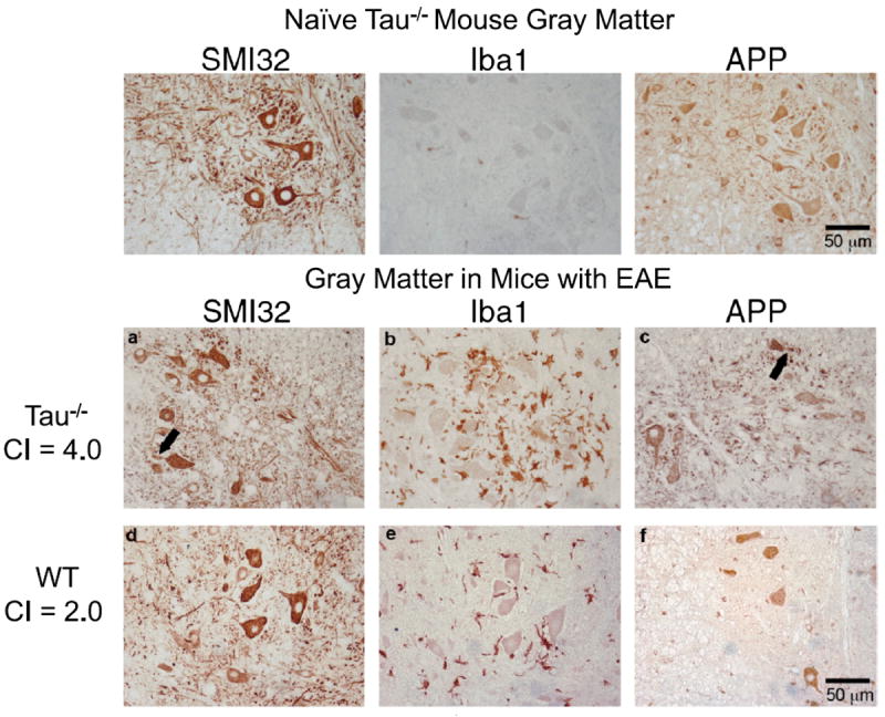
FIGURE 5.

Neuronal damage in gray matter of tau-deficient (Tau−/−) mice in acute experimental autoimmune encephalomyelitis (EAE). Paraffin-embedded sections of lumbar spinal cord from a naive Tau−/− mouse (upper row), a Tau−/− mouse with EAE (day 19) (middle row), and a wild-type (WT) mouse with EAE (day 19) (bottom row) were immunostained with antibodies to amyloid precursor protein (APP), SMI32 and Iba1. (a–c) In Tau−/− mice, neurons with SMI32-positive (a) and APP-positive (c) aberrant neurites (arrows) were near Iba1-positive activated microglia (b). (d–f) The WT mouse with EAE has no detectable SMI32-positive (d) or APP-positive (f) swellings and fewer immunostained microglia (e). CI = clinical index.
