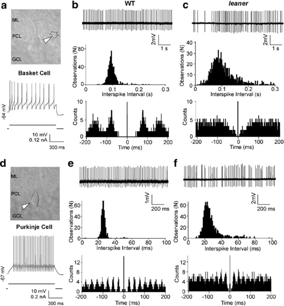
Fig. 4.
Comparison between cell-attached recordings of spontaneous activity in WT and leaner basket and Purkinje neurons. a IR-DIC image of a basket neuron (white arrowhead) with adjacent PC in a WT cerebellar slice. ML molecular layer, PCL Purkinje cell layer, GCL granular cell layer. Recording below shows evoked firing under whole cell current-clamp conditions, illustrating spike frequency adaptation during depolarizing current pulse. b, c Representative cell-attached patch recordings of spontaneous activity in wild-type and leaner basket neurons, with ISI histograms and autocorrelograms below. Mean firing frequency in leaner molecular layer interneurons is similar to WT cells, but ISI variability is more pronounced as indicated by the broader ISI distribution and less defined autocorrelogram peaks, indicative of increased spike rate variability. d IR-DIC image of PC (top, white arrowhead) with recording of evoked firing activity under whole cell current-clamp (bottom). Unlike basket neurons, PCs respond to steady weak depolarizing current injection with sustained repetitive firing without spike frequency adaptation. e, f Representative cell-attached recordings of spontaneous activity from wild-type and leaner PCs with corresponding ISI histograms and autocorrelograms (below). Note higher discharge rate of PCs compared to basket neurons in both WT (compare b and e) and leaner (compare c and e) mice. Also note that cell-attached recordings from WT and leaner PCs confirm differences in firing rate and ISI variability observed during whole cell recordings (see Fig. 1 and ESM Fig. 1)

