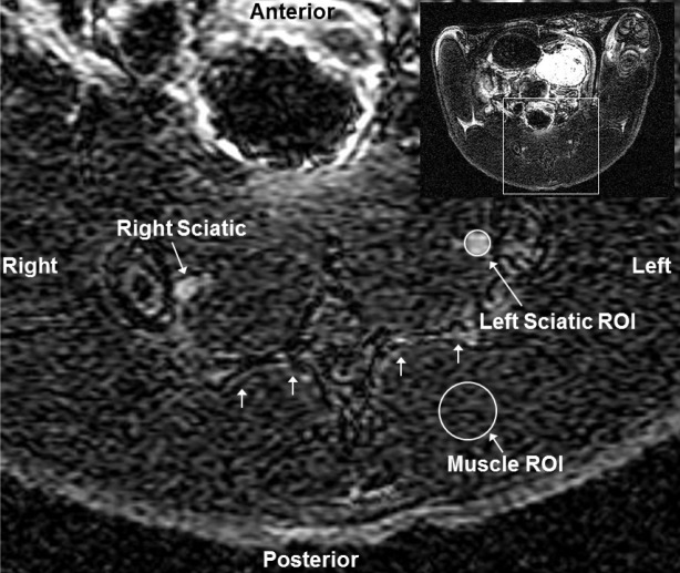
Figure 1.
Placement of ROIs around sciatic nerves. Axial T1 weighted MRI slice of the posterior rat pelvis (inset image of entire transaxial slice shown in upper right corner) showing sacrum (multiple small arrows), right and left sciatic nerves, representative regions-of-interest (ROI) around the left sciatic nerve and muscle. Inset shows the entire slice from which the magnified slice was obtained.

