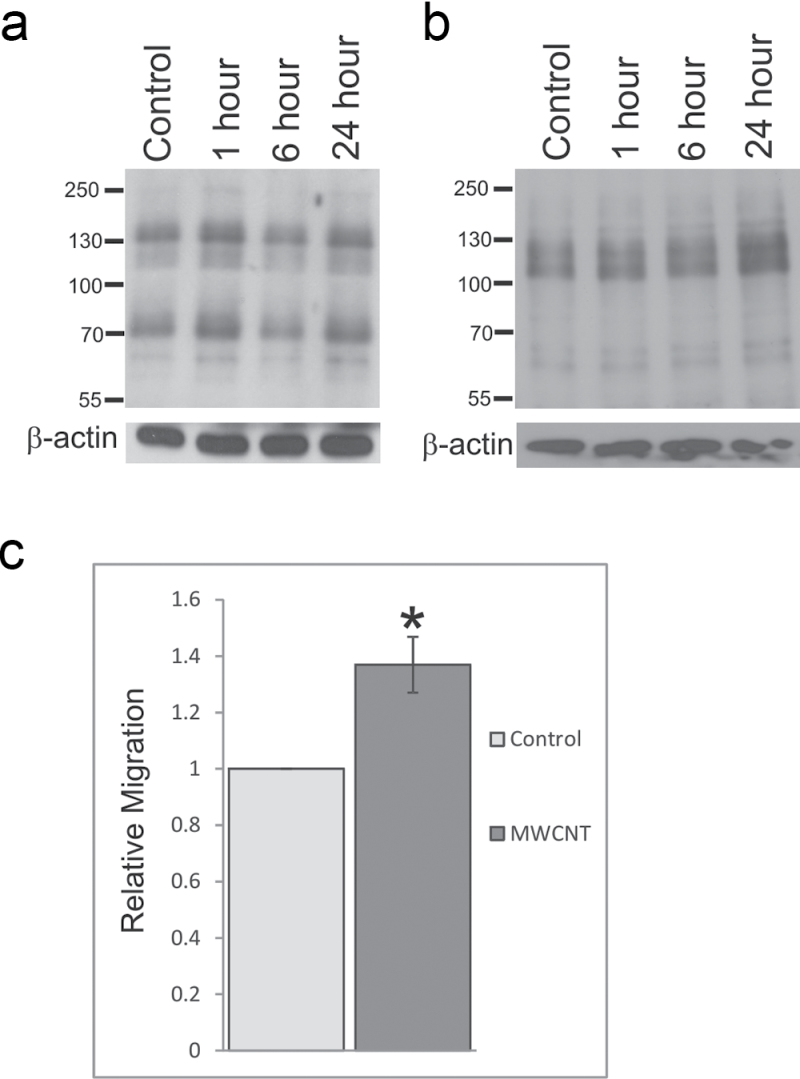
Fig. 3.
MWCNT induces epithelial cell activation. SAEC were serum starved, exposed to DM control or 1.2 µg/ml MWCNT for 1, 6, or 24h, and lysed for analysis by SDS-PAGE. Whole-cell lysates were resolved on an 8% polyacrylamide gel and probed for (a) phosphotyrosine or (b) phosphothreonine. β-Actin was used as a loading control. (c) SAEC migration after MWCNT exposure was determined using a modified Boyden chamber. Migrated cells were stained with 0.1% crystal violet, eluted with acetic acid, and signal intensity read on a microplate reader. Data are presented as mean ± SEM of five assays. * Statistically significant at p ≤ 0.05.

