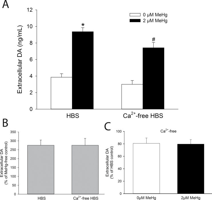
Fig. 2.
Role of extracellular Ca2+ in MeHg-induced DA release. (A) The concentration of extracellular DA was measured from PC12 cells treated for 60min with 0µM (white bars) or 2µM (black bars) MeHg in the presence (HBS) or absence (Ca2+-free HBS) of extracellular Ca2+. * indicates a significant difference from HBS-Control (p ≤ 0.05). # indicates a significant difference from both HBS-Control and HBS-MeHg (p ≤ 0.05). (B) The percentage change of extracellular DA after treatment with MeHg was calculated in cells treated with either HBS or Ca2+-free HBS. (C) The percentage reduction of extracellular DA by treatment with Ca2+-free HBS in cells treated with either 0 or 2µM MeHg. Values are means ± SEM (n = 4, three replicates per n).

