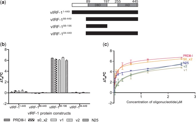
Figure 1.
(a) The predicted domain boundaries of vIRF-1: DBD and the IAD (30). Below are the different domain boundary truncations of our four vIRF-1 protein constructs. (b) A TSSA was performed for all the four vIRF-1 protein constructs with several dsDNA, including the PRDIII-I sequence from the human IRF operator (PRDIII-I), a two times repeat of S0 (S0_x2), two sequences from the viral operator region that contains sequences that are highly similar to S0 (v1 and v2) and a random DNA sequence that contains 25 bp (N25). The TSSA shows that only vIRF-188–196 is stabilized by the various dsDNA. (c) A dose–response experiment results of vIRF-188–196 with varying concentrations of the aforementioned dsDNA. A dose–responsive stabilization of vIRF-188–196 by the different dsDNA was observed.

