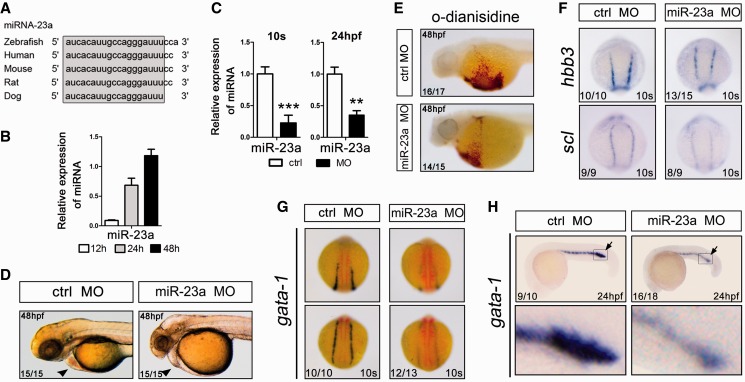
Figure 6.
Suppression of miR-23a blocks erythroid differentiation in zebrafish. (A) Manual alignment of mature miR-23a genomic sequences in five vertebrate species. Sequences derived from the miRBase database (Release 18). (B) qPCR analysis of miR-23a at 12, 24 and 48 h of zebrafish embryo development. Error bars represent standard deviation obtained from three independent experiments. (C) qPCR analysis of miR-23a in 10 somites and 24 hpf embryos after the indicated treatments. (*) Significant changes in embryos injected with the morpholino antagonist of miR-23a (miR-23a MO) compared with control morpholino (Ctrl MO) (*P < 0.05; **P < 0.01). (D) Lateral view of the heart and yolk sac of Ctrl MO- or miR-23a MO-injected embryos. (E) o-Dianisidine staining of hemoglobin in randomly selected 48 hpf embryos injected with Ctrl MO or miR-23a MO. (F) Expression of the hematopoietic lineage markers hbbe3 and scl in 10 somites embryos by whole-mount in situ hybridization. Randomly selected 10 somites embryos, injected with Ctrl MO or miR-23a MO, showing a normal degree of hbbe3 and scl staining in both groups. (G and H) Expression of gata-1 in 10 somites (G) and 24 hpf (H) embryos by whole-mount in situ hybridization. Randomly selected 10 somites and 24 hpf embryos, injected with Ctrl MO or miR-23a MO, showing a normal degree of gata-1 staining in both groups.

