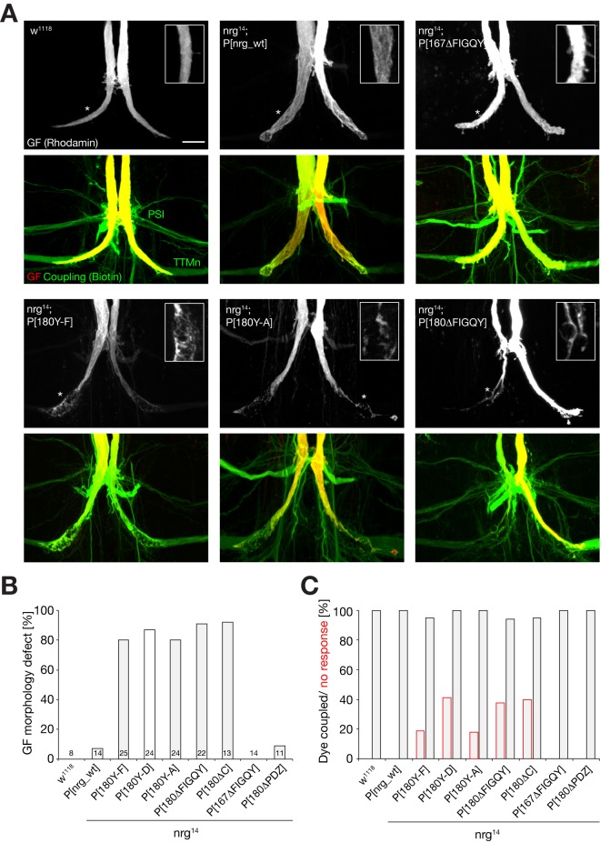
Figure 7. Anatomical phenotypes of the giant fiber synaptic terminals in nrg mutants.
(A) GF synaptic terminals were visualized by injection of Rhodamine-dextran (red) into the GF. Dye-coupling of the GF to its target neurons, the Tergo Trochanteral motoneuron (TTMn), and the peripheral synapsing interneuron (PSI) via co-injection of Biotin (green) allows the detection of gap junctions between these neurons. In w1118, nrg 14; P[nrgwt] and nrg 14; P[nrg167ΔFIGQY], a normal, large GF terminal was present and we observed dye-coupling with the TTMn and the PSI. In nrg 14; P[nrg180Y-F], nrg 14; P[nrg180Y-A], and nrg 14; P[nrg180ΔFIGQY] mutants, the presynaptic terminal of the GF exhibited variable abnormal morphologies. They were thinner or swollen and contained large vacuole-like structures. However, in most cases the GF still dye-coupled with the postsynaptic target, the TTMn, and the PSI. Scale bar, 15 µm. (B) Quantification of morphological defects in w1118 flies and nrg mutants. Only mutations affecting the Nrg180–FIGQY motif resulted in severe GF terminal aberrations. (C) Quantification of GF-to-TTMn dye-coupling (black bars) and comparison to animals with no electrophysiological responses (red bars) of the TTM with GF stimulation in the brain. A large percentage of animals expressing mutant versions of Nrg180–FIGQY proteins completely lacked electrophysiological responses despite the presence of dye-coupling, demonstrating a severe functional defect in these animals.

