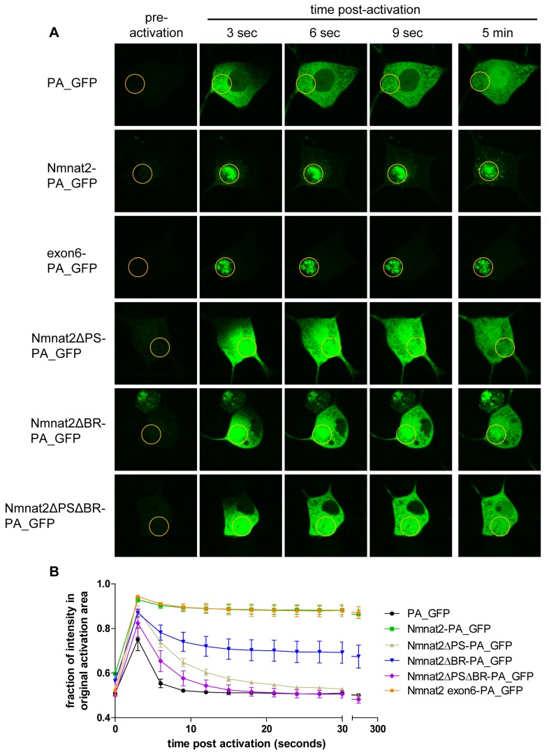
Figure 2. Exon 6 sequences are necessary and sufficient for Nmnat2 membrane association.
(A) Use of photoactivatable GFP (PA_GFP) fusion proteins to study Nmnat2 membrane association. Time course of a representative cell body of an SCG primary culture neuron injected with each indicated construct is shown. The region of activation is marked by an orange circle in each image. (B) Quantification of protein mobility. Shown is the percentage of fluorescence that remains in the originally activated area compared to a non activated area of equal size elsewhere in the cell body. Error bars indicate SEM.

