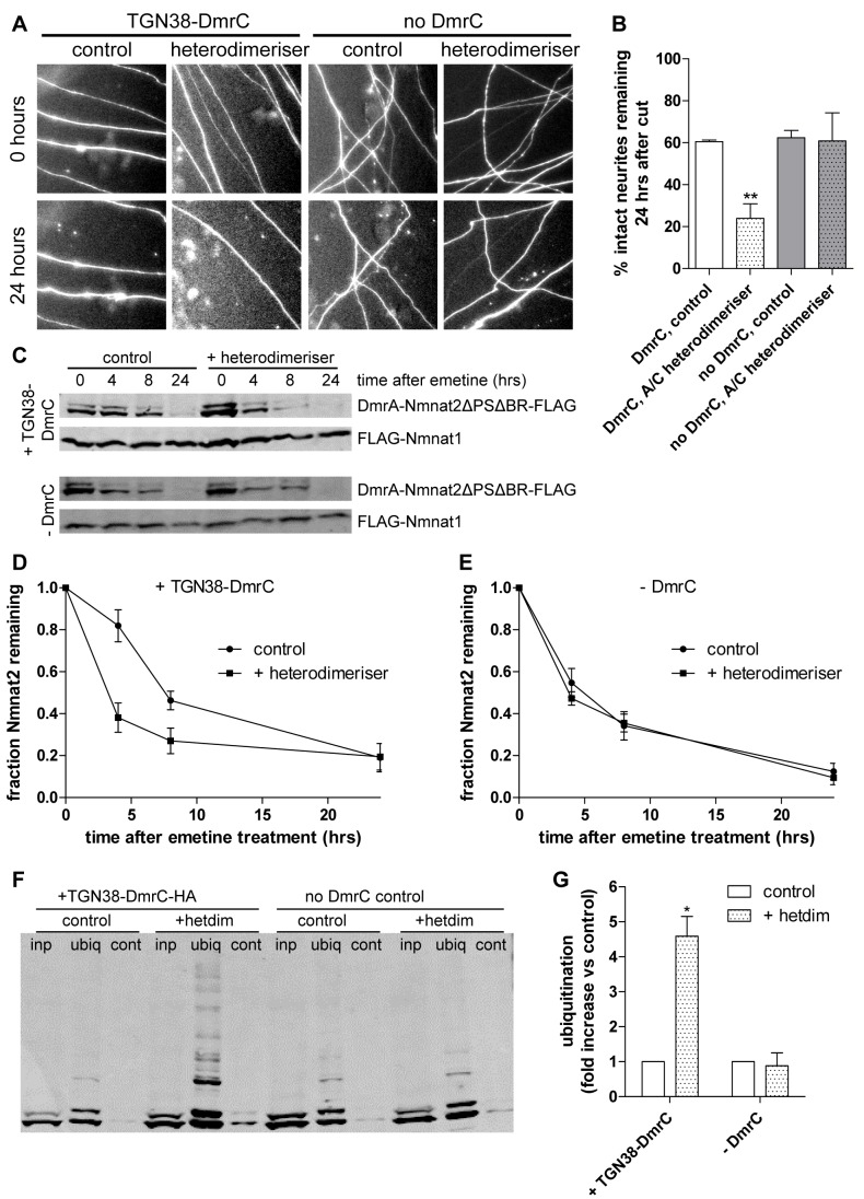
Figure 8. Membrane re-targeting through heterodimerisation causes ubiquitination, reduced protein stability, and impaired neurite protection by Nmnat2ΔPSΔBR.
(A) Representative fields of view of distal primary culture SCG neurites 0 and 24 h after neurite cut, labelled by dsRed2 fluorescence and injected with 0.002 µg/µl of DmrA-Nmnat2ΔPSΔBR-PA_GFP plus 0.01 µg/µl TGN38-DmrC-HA or 0.001 µg/µl of DmrA-Nmnat2ΔPSΔBR-PA_GFP without any TGN38-DmrC-HA present. Eight hours before neurite cut, 500 nM of A/C heterodimeriser was added to relevant cultures. (B) Quantification of experiment shown in (A). Error bars indicate SEM. ** indicates statistically significant difference compared to control (** p<0.01). (C) Representative Western blots of HEK293 cells co-transfected with DmrA-Nmnat2ΔPSΔBR-FLAG, FLAG-Nmnat1, and TGN38-DmrC-HA (+TGN38-DmrC) or empty pCMV-Tag4A vector (-DmrC). To induce heterodimerisation, 500 µM A/C heterodimeriser were added to the appropriate wells. Twenty-four hours after transfection, cells were treated with 10 µM emetine for the amount of time indicated, after which samples were processed for SDS-PAGE and Western blot using anti-FLAG antibody. (D) Quantification of DmrA-Nmnat2ΔPSΔBR-FLAG turnover after emetine treatment when co-transfected with TGN38-DmrC-HA. For each sample and time point, the amount of DmrA-Nmnat2ΔPSΔBR-FLAG remaining was normalised to FLAG-Nmnat1 as an internal control. Error bars indicate SEM. (E) Quantification of DmrA-Nmnat2ΔPSΔBR-FLAG turnover after emetine treatment in the absence of TGN38-DmrC-HA. For each sample and time point, the amount of DmrA-Nmnat2ΔPSΔBR-FLAG remaining was normalised to FLAG-Nmnat1 as an internal control. Error bars indicate SEM. (F) Representative Western blot of GST-Dsk2 pulldown assay. HEK293 cells expressing DmrA-Nmnat2ΔPSΔBR-FLAG and TGN38-DmrC-HA and maintained in the absence (control) or presence (+heterodimeriser) of 500 µM A/C heterodimeriser were lysed (inp – total input), and ubiquitinated proteins were immunoprecipitated using GST-Dsk2 bound to glutathione beads (ubiq). GST-fused mutant Dsk2 was used for control pulldown (cont). Eluted proteins were processed for SDS-PAGE and analysed by Western blot using anti-FLAG antibody. (G) Quantification of ubiquitination assay. For each condition, the total amount of ubiquitinated DmrA-Nmnat2ΔPSΔBR-FLAG was normalised to total input. Error bars indicate SEM. *** indicates statistically significant difference compared to control (*** p<0.001).

