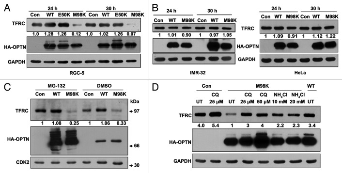
Figure 3. M98K expression induces TFRC degradation through lysosomal pathway in RGC-5 cells. (A) Western blot shows TFRC levels in RGC-5 cells infected with control (con), WT, E50K or M98K adenoviruses after 24 and 30 h of expression. GAPDH was used as a loading control. The numbers below TFRC blot indicate relative TFRC levels after normalization. (B) Effect of M98K expression on TFRC levels in IMR-32 (left panel) and HeLa (right panel) cells. (C) M98K-mediated TFRC degradation is independent of proteasomal degradation. RGC-5 cells infected with control (con), WT or M98K adenoviruses for 12 h were treated with either DMSO or with 5 μM MG-132 for further 12 h. Cell lysates were then subjected to western blotting. CDK2 was used as a loading control. (D) Effect of lysosomal inhibitors on M98K-mediated TFRC degradation. After 6 h of infection with the indicated adenoviruses, RGC-5 cells were either left untreated or treated with 25 and 50 μM of chloroquine or 10 and 20 mM of ammonium chloride for 24 h. Whole cell lysates were subjected to western blotting. CQ, chloroquine.

An official website of the United States government
Here's how you know
Official websites use .gov
A
.gov website belongs to an official
government organization in the United States.
Secure .gov websites use HTTPS
A lock (
) or https:// means you've safely
connected to the .gov website. Share sensitive
information only on official, secure websites.
