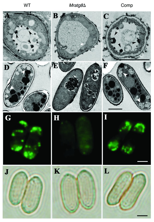
Figure 5.MrATG8 effects on the accumulation of autophagosome and lipid droplets (LDs). After growth in an MM-N medium for 4 h, vacuolar autophagic bodies (arrows) were evident in the WT (A) and Comp (C) cells but not in the gene-deletion mutant (B). Conidia in the wild type (WT), Mratg8Δ and Comp mutant harvested after 20 d on CM plates were used for TEM analysis. In contrast to the WT (D) and Comp (F), accumulation of LDs (arrows) was significantly reduced in Mratg8Δ (E). This was confirmed by Bodipy fluorescent dye staining that shows the lack or presence of a few LDs in Mratg8Δ conidia (comparing H to G and I). (J–L) show the corresponding bright-field images of (G–I), respectively. Scale bar: 2 μm.

An official website of the United States government
Here's how you know
Official websites use .gov
A
.gov website belongs to an official
government organization in the United States.
Secure .gov websites use HTTPS
A lock (
) or https:// means you've safely
connected to the .gov website. Share sensitive
information only on official, secure websites.
