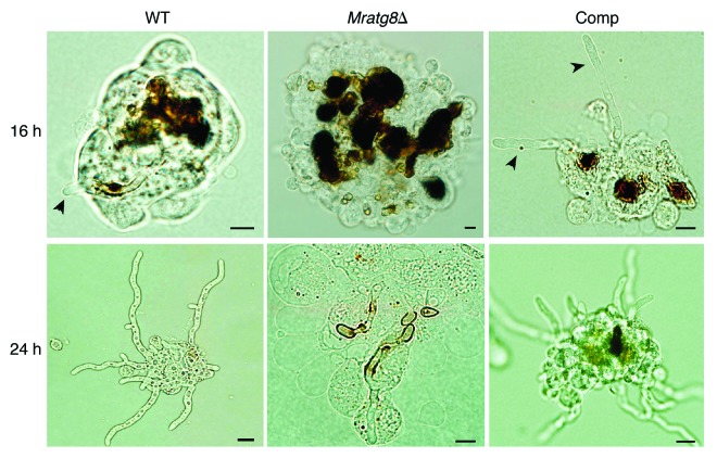
Figure 9. Fungal development in insect hemocoel. Relative to the WT and Comp, Mratg8Δ conidia were heavily encapsulated and melanized by insect hemocytes 16 h after injection. Arrows indicate the escaped hyphal tubes. Twenty-four hours postinjection, mycelia of the WT and Comp branched and escaped from the hemocyte nodules but not in the gene-deletion mutant. Scale bar: 10 µm.

An official website of the United States government
Here's how you know
Official websites use .gov
A
.gov website belongs to an official
government organization in the United States.
Secure .gov websites use HTTPS
A lock (
) or https:// means you've safely
connected to the .gov website. Share sensitive
information only on official, secure websites.
